Watch: This Time-Lapse Boldly Goes Where No Time-Lapse Has Gone Before
Watching a time-lapse of a sunset or the construction of a skyscraper is cool, but what about the creation of life?
Time-lapse videos are all over the place, and there are definitely some that are out-of-this-world amazing. You probably have your favorites: the Calbulco volcanic eruption, Architect by Kevin McGloughlin, or, my personal favorite, the one of Singapore that took 3 years to shoot. However, sometimes it's not so much about the great cinematography or creative use of mixed media. Sometimes it's just about capturing something that represents something important to the human existence, like the very first moments of life—of a tadpole.
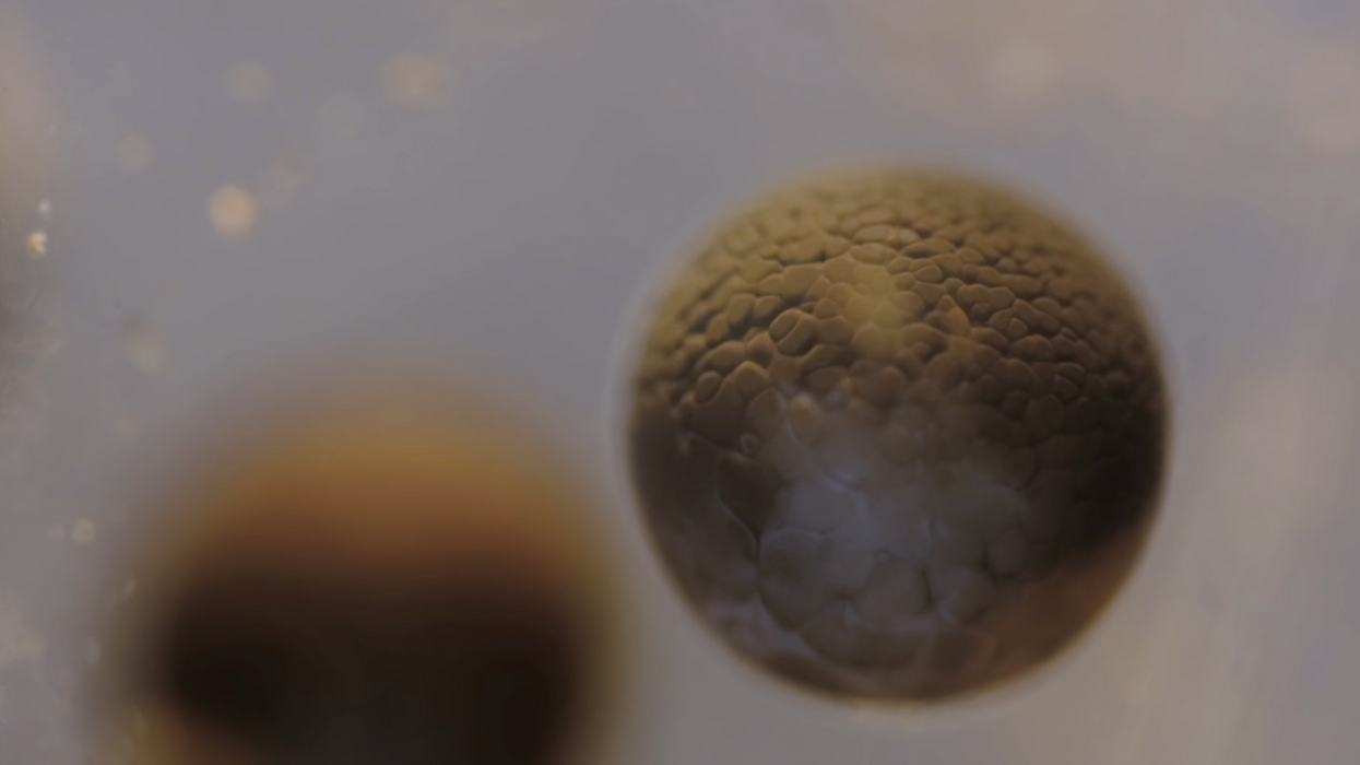
This incredible time-lapse video of the cell division of a Rana temporaria egg, better known as the common frog, was captured by filmmaker Francis Chee. Though it only lasts 23 seconds, it took 33 hours to record. According to the video's description, Chee used a custom designed microscope he built himself that was "based on the infinity optical design." He also made customized LED lights and optics to illuminate the egg. He explains his process a little more in depth:
The whole microscope sits on anti-vibration table. I have to say that it doesn't matter too much what microscope people use to perform this. There are countless other variables involved in performing this tricky shot, such as for example: the ambient temperature during shooting, the time at which the eggs were collected, the handling skills of the operator, the type of water used, lenses, quality of camera, etc.
A video like this is incredible because, well, because it's real. I mean, we've all seen those gritty black and white videos in science class that show us the wonderful bore that is mitosis, but Chee's video looks like it was made by some VFX artist. But it wasn't! That's actually what cell division looks like. It's real! It's science! It's filmmaking! It's real science captured by filmmaking and that's exciting, guys!
Source: Sploid